Could the invention of ‘molecular GPS’ help us develop better medicines?
Scientists have developed a new method to locate exactly where a drug interacts with proteins in our cells.
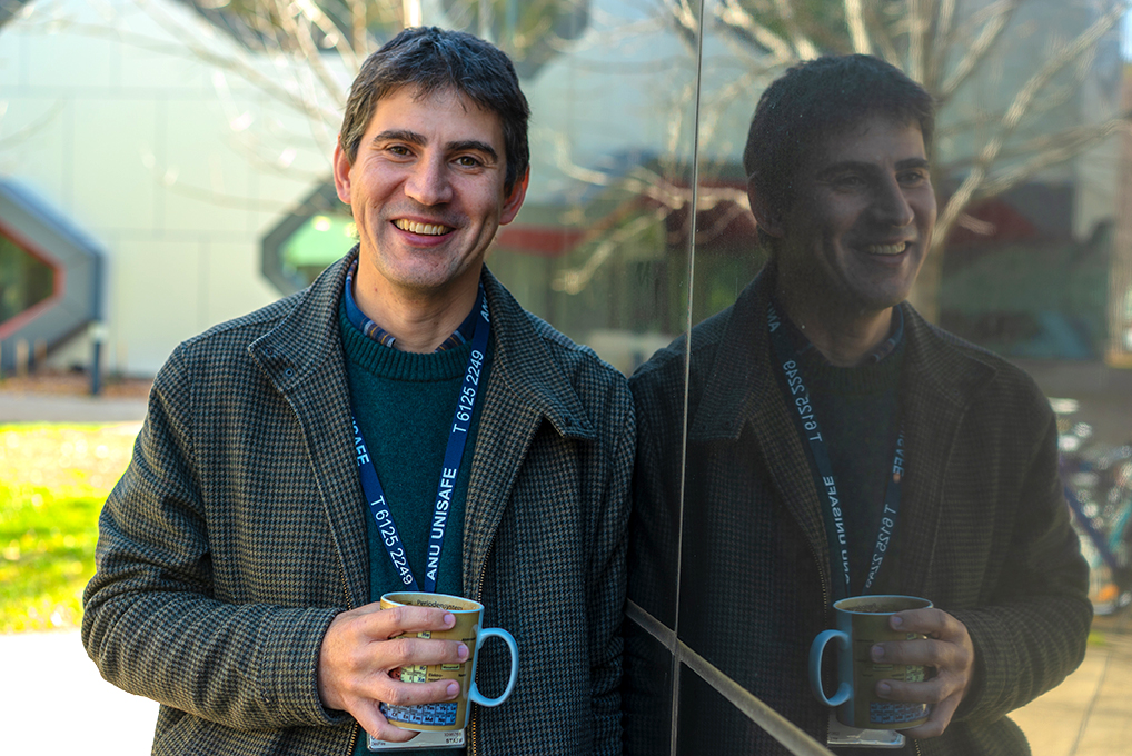
“Just like how GPS triangulates your location using satellites, we can place labels at different positions around the surface of a protein to triangulate the position of a fluorinated molecule within it,” says Dr Martyna Judd.
The new method offers a unique solution for visualising drug-binding interactions that are difficult to see with current conventional methods.
“When developing a drug for the market, it’s essential to understand how it interacts with the target protein,” says Dr Judd.
“This is critical to evaluating drug efficacy, optimising its design, and preventing unwanted side effects.”
Dr Judd is a co-author of the paper published in the Journal of the American Chemical Society (JACS) and completed her research at The Australian National University (ANU) Research School of Chemistry.
“Having a molecular-level lens into protein-drug interactions is critical in scenarios like antimicrobial resistance,” she explains.
“Especially where even small mutations in the target enzyme structure can completely change a drug’s mode of activity, rendering it inactive.”
The new, more sensitive technique, developed in collaboration with researchers from Germany and Australia, uses magnetically active molecules as ‘labels’ on a protein surface.
“The magnetic labels can help us detect nearby fluorine atoms present in interacting molecules, such as drug molecules binding to the protein.”
Conveniently, over 20 per cent of today’s pharmaceuticals contain a fluorine atom in their structure.
Another limiting factor to current conventional methods is the distance range across which they can detect a drug molecule.
But by triangulating the distances to the fluorine sites from multiple different locations, Dr Judd says the new method “improves the robustness of the picture we are trying to reconstruct”.
The technology has real potential for researchers to visualise how drugs are transported across cell membranes in the future.
“Interaction with membrane proteins is a critical determinant of a drug’s bioavailability,” she says.
“With further improvements to the magnetic tags and detection strategies currently underway by our team and other research groups, we are getting close to measuring distances approaching almost half the width of a biological membrane.
“You could one day imagine installing a magnetic label on either side of a cell membrane to potentially monitor both the entry and exit of a fluorinated drug through the cell wall.”
This new method is based on electron paramagnetic resonance spectroscopy (EPR), which uses magnetic fields and microwave radiation to study materials with unpaired electrons.

ANU is home to the only high-field EPR facility in the Southern Hemisphere. The facility is led by Professor Nick Cox, and it has wide-ranging applications for medical, biological, chemical and materials research.
Working with such advanced technology was a highlight of Dr Judd’s PhD journey at ANU.
“Not only did I get hands-on experience on unique, advanced machinery, but I also had the opportunity to engage in very interesting work on samples from highly interdisciplinary collaborations.”
Dr Judd recently started a position as a postdoctoral researcher at Northwestern University, but her chemistry journey began at ANU, where she also completed her undergraduate Bachelor of Philosophy (Honours) degree. Her interest in spectroscopy was sparked when she completed a course led by her PhD supervisor, Professor Nick Cox.
“I became fascinated with the puzzle of working out molecular structures from different, individual pieces of experimental information.”
This passion eventually led to a Fulbright Scholarship, allowing her to travel to the US during her PhD to work at Northwestern University, fine-tuning a different type of spectroscopy.
“Keeping in the theme of fluorine measurements for biological systems, my project focused on building a specialised probe made of fluorine-free materials, for directly detecting fluorines with enhanced signal sensitivity enabled by microwave irradiation.”
At Northwestern University, Dr Judd is now continuing her work on fluorine detection and expanding into new areas of molecular spectroscopy.
Back in Australia, her collaborators are putting the new ‘molecular GPS’ method to the test by investigating a new anti-inflammatory drug target.
Thanks to an ARC Linkage Program grant, Professor Ross Bathgate from the Florey Institute and Professor Paul Gooley from the University of Melbourne are investigating the pregnancy hormone relaxin as a potential new therapeutic without the severe side effects of current options.
Dr Judd is hopeful that the new method will be used many more times to understand how a wide range of drugs work in our bodies.
“Developing new therapeutics revolves around targeting proteins or enzymes, and EPR offers measurement sensitivity unparalleled by other methods, allowing us to access and visualise drug-protein interactions inside the actual cell environment.”
It turns out that achieving greater precision in drug design could be all about accurately tracking where new drugs go.
This study was funded by the ARC Discovery Program and is a collaboration between researchers at the ANU, Bielefeld University in Germany, Dortmund University in Germany, the University of Queensland and the Australian Research Council Centre of Excellence in Protein and Peptide Science.


